Beranda
/ Anatomy Of Chest / Chest Wall Anatomy Springerlink - The epidermis is the outermost layer that provides a protective, waterproof seal over the body.
Anatomy Of Chest / Chest Wall Anatomy Springerlink - The epidermis is the outermost layer that provides a protective, waterproof seal over the body.
Insurance Gas/Electricity Loans Mortgage Attorney Lawyer Donate Conference Call Degree Credit Treatment Software Classes Recovery Trading Rehab Hosting Transfer Cord Blood Claim compensation mesothelioma mesothelioma attorney Houston car accident lawyer moreno valley can you sue a doctor for wrong diagnosis doctorate in security top online doctoral programs in business educational leadership doctoral programs online car accident doctor atlanta car accident doctor atlanta accident attorney rancho Cucamonga truck accident attorney san Antonio ONLINE BUSINESS DEGREE PROGRAMS ACCREDITED online accredited psychology degree masters degree in human resources online public administration masters degree online bitcoin merchant account bitcoin merchant services compare car insurance auto insurance troy mi seo explanation digital marketing degree floridaseo company fitness showrooms stamfordct how to work more efficiently seowordpress tips meaning of seo what is an seo what does an seo do what seo stands for best seotips google seo advice seo steps, The secure cloud-based platform for smart service delivery. Safelink is used by legal, professional and financial services to protect sensitive information, accelerate business processes and increase productivity. Use Safelink to collaborate securely with clients, colleagues and external parties. Safelink has a menu of workspace types with advanced features for dispute resolution, running deals and customised client portal creation. All data is encrypted (at rest and in transit and you retain your own encryption keys. Our titan security framework ensures your data is secure and you even have the option to choose your own data location from Channel Islands, London (UK), Dublin (EU), Australia.
Anatomy Of Chest / Chest Wall Anatomy Springerlink - The epidermis is the outermost layer that provides a protective, waterproof seal over the body.. Here, we break down the anatomy of your chest muscles. This atlas is a comprehensive and affordable learning tool for medical students and residents and especially for radiologists and pneumologists. Radiology basics of chest ct anatomy with annotated coronal images and scrollable axial images to help medical students and junior doctors learning anatomy. See human chest anatomy stock video clips. Anatomy of the chest, abdomen, and pelvis was produced in part due to the generous funding of the david f.
You will also find the xiphoid process, 10th rib, the apex of the heart, the coronary vein of the heart. Anatomy of the eye 12 photos of the anatomy of the eye anatomy of the eye bones, anatomy of the eye lacrimal gland, anatomy of the eye special senses vision, anatomy of the eye ultrasound, external anatomy of the eye quiz, human anatomy, anatomy of the eye bones, anatomy of the eye lacrimal gland, anatomy … And flexibility to aid in the functional process of respiration. Organs & structures of the chest heart. A typical heart is approximately the size of your fist:
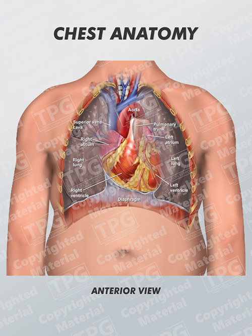
Chest Anatomy Order from presentationgroup.com Stability to arm and shoulder movement; See human chest anatomy stock video clips. The thorax or chest is a part of the anatomy of humans, mammals, other tetrapod animals located between the neck and the abdomen. The chest is made up primarily of two muscles: Related posts of anatomy of the chest area anatomy of rib cage. Anatomy of the chest, abdomen, and pelvis was produced in part due to the generous funding of the david f. Swensen fund for innovation in teaching. Anatomy of the thorax, heart, abdomen and pelvis recommended text gray's anatomy for students.
Table 1.1 lists the major anatomic structures within the thorax that are discussed.
Computed tomography (ct) of the chest can detect pathology that may not show up on a conventional chest radiograph(1). Here, we break down the anatomy of your chest muscles. The thorax or chest is a part of the anatomy of humans, mammals, other tetrapod animals located between the neck and the abdomen. Related posts of anatomy of the chest area anatomy of rib cage. Of the two chest muscles, the pectoralis major (a.k.a. The muscles of the chest develop from the somites found in the mesoderm. A good radiologist knows the anatomy because knowing where structures normally live and recognizing the location of an abnormality helps to make or narrow the differential diagnosis. Basic thoracic anatomy and physiology an understanding of thoracic imaging requires knowledge of the anatomy being imaged, as described in this chapter, as well as the imaging techniques applied to the thorax, covered in chapter 2. You will also find the xiphoid process, 10th rib, the apex of the heart, the coronary vein of the heart. However, the classical anatomical descriptions in textbooks make it difficult to gain full mastery of this subject, because the books usually deal with its elements separately. The chest is the area of origin for many of the body's systems as it houses organs such as the heart, esophagus, trachea, lungs, and thoracic diaphragm. Chest bone, ribs, lung, heart, xiphoid process, sternum anatomy. Stability to arm and shoulder movement;
The muscles of the chest develop from the somites found in the mesoderm. An overview of the anatomy visible in a transverse computed axial tomographical image of the thorax (and part of the abdomen) performed with intravenous cont. This atlas is a comprehensive and affordable learning tool for medical students and residents and especially for radiologists and pneumologists. Of the two chest muscles, the pectoralis major (a.k.a. 12 cm (5 in) in length, 8 cm (3.5 in) wide, and 6 cm (2.5 in) in thickness.
Human Chest Anatomy Diagram Koibana Info Anatomy Human Anatomy Body Anatomy from i.pinimg.com Anatomy of rib cage 12 photos of the anatomy of rib cage anatomical rib cage necklace, anatomy and. Organs & structures of the chest heart. First i'll do an intro to the different organs and structures in the chest, and then i'll go over some images showing their locations. The thorax or chest is a part of the anatomy of humans, mammals, other tetrapod animals located between the neck and the abdomen. This page provides an overview of the chest muscle group. A typical heart is approximately the size of your fist: Computed tomography (ct) of the chest can detect pathology that may not show up on a conventional chest radiograph(1). Anatomy of the thorax, heart, abdomen and pelvis recommended text gray's anatomy for students.
The pec major) is the one that commands the most real estate.
See human chest anatomy stock video clips. The epidermis is the outermost layer that provides a protective, waterproof seal over the body. Anatomically, the heart is located in the anterior thoracic cavity; Thoracic cavity, also called chest cavity, the second largest hollow space of the body. (1) the pectoralis major, and (2) the pectoralis minor. However, the classical anatomical descriptions in textbooks make it difficult to gain full mastery of this subject, because the books usually deal with its elements separately. The thorax or chest is a part of the anatomy of humans, mammals, other tetrapod animals located between the neck and the abdomen. First i'll do an intro to the different organs and structures in the chest, and then i'll go over some images showing their locations. Three dimensional view of the female reproductive system, full frontal view. The chest anatomy includes the pectoralis major, pectoralis minor and the serratus anterior. The muscles of the chest develop from the somites found in the mesoderm. The chest is the area of origin for many of the body's systems as it houses organs such as the heart, esophagus, trachea, lungs, and thoracic diaphragm. It is enclosed by the ribs, the vertebral column, and the sternum, or breastbone, and is separated from the abdominal cavity (the body's largest hollow space) by a muscular and membranous partition, the diaphragm.
About the 6th week, the somites differentiate into the sclerotomes and the dermatomyotomes. Of the two chest muscles, the pectoralis major (a.k.a. The chest or thorax is the region between the neck and diaphragm that encloses organs, such as the heart, lungs, esophagus, trachea, and thoracic diaphragm. A good radiologist knows the anatomy because knowing where structures normally live and recognizing the location of an abnormality helps to make or narrow the differential diagnosis. A typical heart is approximately the size of your fist:
Thoracic Cavity Description Anatomy Physiology Britannica from cdn.britannica.com 12 cm (5 in) in length, 8 cm (3.5 in) wide, and 6 cm (2.5 in) in thickness. This page provides an overview of the chest muscle group. The anatomic illustrations are presented as… The muscles of the chest develop from the somites found in the mesoderm. First i'll do an intro to the different organs and structures in the chest, and then i'll go over some images showing their locations. The chest wall is a complex system that provides rigid protection to the vital organs such as the heart, lungs, and liver; It provides access to ct images in the axial plane, allowing the user to learn and review the lung anatomy interactively. Anatomically, the heart is located in the anterior thoracic cavity;
Stability to arm and shoulder movement;
The anatomic illustrations are presented as… Anatomy of the chest, abdomen, and pelvis was produced in part due to the generous funding of the david f. And flexibility to aid in the functional process of respiration. How to view the anatomical labels. Anatomy of the thorax, heart, abdomen and pelvis recommended text gray's anatomy for students. It provides access to ct images in the axial plane, allowing the user to learn and review the lung anatomy interactively. See human chest anatomy stock video clips. Anatomy of the eye 12 photos of the anatomy of the eye anatomy of the eye bones, anatomy of the eye lacrimal gland, anatomy of the eye special senses vision, anatomy of the eye ultrasound, external anatomy of the eye quiz, human anatomy, anatomy of the eye bones, anatomy of the eye lacrimal gland, anatomy … Anatomically, the heart is located in the anterior thoracic cavity; 12 cm (5 in) in length, 8 cm (3.5 in) wide, and 6 cm (2.5 in) in thickness. A typical heart is approximately the size of your fist: (1) the pectoralis major, and (2) the pectoralis minor. Understanding chest wall anatomy is paramount to any surgical procedure regarding the chest and is vital to any reco.

